Dental Portal presents an updated review on fitting dental crowns. From ceramic to zirconia, we explore various types of crowns and their benefits. Learn how to preserve the nerve and the techniques used for placing crowns on a post or in an inlay. We also discuss how to replace an old crown and how to prepare for your first visit to the dental office. Gain all the necessary information to make an informed decision about your treatment.
Welcome to Dental Portal! In our latest article, we delve into all aspects of fitting dental crowns. Discover how to select the perfect material and understand the process from start to finish. Your journey to a perfect smile begins here!
What is a Dental Crown?

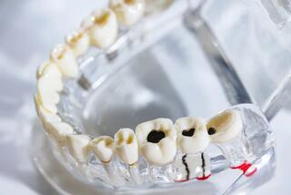
A dental crown is an orthopedic prosthesis, placed on a preserved root or implant to restore the anatomical shape of a tooth. An artificial crown is necessary when it's impossible to use a filling or veneers.
We restore the beauty of your smile with prostheses – selecting a color that matches the natural shade of your enamel. After the crown placement, the transition area becomes imperceptible, and a beautiful gingival margin is restored.
Dental Crowns - Before and After
A clinical case – teeth were destroyed, the patient experienced constant discomfort. The diagnosis was bruxism.
The treatment result – a complete reconstruction of both the upper and lower jaws was conducted. The bite was restored, bruxism was eliminated, and then zirconia crowns were placed on the preserved teeth.
Types of Dental Crowns by Material
Ceramic
Crowns made of ceramic are created without a metal framework – this is the best option for prosthetics of front teeth. The ceramic framework fully matches the color of the dental row, and the surface mimics the semi-transparency of enamel.
Ceramic crowns IPS E-max are suitable for individual restorations or for bridging in the frontal section. The structure is impeccably aesthetic and fully biocompatible.
Zirconia
Zirconia crowns are extremely durable, suitable for restoring both front and chewing teeth. The structure withstands high masticatory load.
We create crowns from zirconium dioxide:
- Solid Zirconia – fabricated from a monolithic zirconia block. It's exceptionally strong, enduring a significant load. Ideal for individual restorations on molars or for bridge prosthetics.
- Multi-layer Zirconia – we use multi-layered zirconium dioxide with a gradient color matching enamel. Recommended for prosthetics in the frontal jaw section.
Which material should you choose?
Doctors recommend restoring front teeth with ceramic E-max crowns, as they best match the color of enamel.
For the restoration of side chewing teeth, zirconia crowns are preferred. They are the most durable to withstand masticatory pressure. For hyperfunction of masticatory muscles and bruxism, products with a metal framework are advised.
How to Place a Crown While Preserving the Nerve
If the damage is minor and the pulp is healthy, it is preserved. In other cases, at least part of the living tissue is saved through vital pulpotomy. Then, nutrients continue to flow, dentin is renewed, sensitivity and strong structure are maintained. This prevents further tissue destruction and extends the lifespan of the construction. Prosthetics are possible with quality anesthesia, cooling, and the use of special tips for grinding.
Placing a Crown on a Post
Restoring a tooth on a metal post can lead to its loss due to the wedging effect of the construction.
If the volume and height of tissues are insufficient for prosthetics, they are replenished with a fiberglass post. Its structure, rigidity, and strength resemble dentin, hence the masticatory load is evenly distributed, and internal factors for root destruction do not occur.
The post is set into previously treated channels using an adhesive composition. For creating an artificial core, composite is applied in layers. This structure helps to support the prosthesis, providing additional reinforcement.
Placing a Crown on an Inlay
If the supragingival part is completely destroyed or weakened, but the root can withstand masticatory pressure, the crown is placed on a core build-up inlay. It consists of primary and additional posts inserted into the root canals, and an artificial core.
The inlay is fabricated based on individual impressions. The posts replicate the shape of the tooth's canals and are fixed with composite cement after initial canal filling. Thus, the structure is hermetic, stable, and strong.
The inlay can be monolithic or sectional. A single-piece construction is used for incisors and canines with 1–2 roots, while a sectional one is for premolars and molars with 3–4 roots.
The materials of the inlay and the crown should be compatible. For a prosthesis made of pressed ceramic or zirconium dioxide, a metal-free core build-up inlay is suitable.
How to Place a Crown on a Tooth That Had One Previously Placed
If the prosthesis needs to be replaced, it is removed in one of three ways:
- Cut through the center and remove in parts.
- The cement at the base is broken, the prosthesis is shifted and removed using the Koppa device, which resembles forceps.
- The orthopedic construction is removed with the "Coronaflex" device. A shock wave of compressed air precisely destroys the dental cement, allowing the prosthesis to be easily removed. It remains intact: without scratches, cracks, chips, or deformations.
If the crown falls off, only a dentist can decide on its reuse. Reusing the same prosthesis is impossible if it was deformed during removal, or if there is a gap between its inner surface and the core. Bacteria and food particles will enter this space, leading to inflammation and an infectious process.
If the crown has come off, it should be rinsed, kept in a dry, clean place, and a visit to the dentist should be scheduled promptly. The core is weakened, sensitive to food and drinks, and susceptible to cariogenic bacteria, hence it may quickly deteriorate.
For prosthetics, you can choose crowns made of E-max ceramic or zirconium dioxide. They perfectly restore aesthetics, and withstand greater than masticatory load. Their lifespan is 15–20 years, requiring tissue reduction of 0.8–1.2 mm. Metal-ceramic lasts three times less, changes the color of the adjacent gum part, requires tooth reduction of 1.5–2 mm, therefore, the vascular-nerve bundle is necessarily removed.
First Visit for Crown Placement
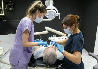
During the first visit, the orthopedic dentist examines the dental-jaw system, prepares the tooth for prosthetics, and makes impressions for the future crown.
-
Diagnosis (10–15 minutes)
Preparation begins with computed tomography of the maxillofacial area. The doctor assesses the condition of the roots, the quality of canal filling, and determines whether a crown can be placed or if another method should be considered. CT scans exclude inflammatory processes, cysts near the root apex, and hidden caries. -
Planning (from 10 minutes to 7 days)
The dentist visually assesses the condition of the oral cavity, evaluates the bite, the degree of destruction of the unit, whether prosthetics are possible with the current volume and height of the crown part, and whether it is necessary to adjust the occlusion of the dental rows. The doctor discusses the stages, duration, cost of treatment, selects the color, and material of the crown.
To prevent the development of caries under the construction and other complications, the entire oral cavity is treated. Soft plaque, dental calculus are removed, cavities with carious are prepared, root canals are treated, and inflammatory diseases of the periodontium are addressed. -
Obtaining the Key (10 minutes)
Using Wax-Up and Mock-Up technologies, the base for making a temporary crown is created. Before grinding, the dentist takes a plaster model, from which a wax model is made. If the patient is satisfied, the dentist prepares a silicone template, which helps to capture the natural relief of the chewing surface and replicate it on the temporary construction. -
Grinding (30–60 minutes)
The dentist removes the surface tissues to the thickness of the future construction. The core is shaped to allow it to be covered by the prosthesis. -
Tooth Depulpation (30 minutes)
If necessary, the dentist performs depulpation: opening the pulp chamber, removing nerves, vessels, connective tissue. Then, the root canals are expanded, cleaned, treated with an antiseptic, and filled with a sealing material – this helps to prevent infection and root destruction. -
Retraction (5 minutes)
To get a clear impression, access to the area where the tooth surface meets the gum is needed. Soft tissues are retracted using cotton thread, a retraction ring, or a silicone cone inserted into the gingival sulcus. -
Taking an Impression (15 minutes)
To ensure the crown fits snugly on the tooth, an impression of the core is made. For making the impression, a pliable mass is used: it is placed in a dental tray resembling a cap, then fixed on the dental row. -
Temporary Crown (15–20 minutes)
The ground tooth is hypersensitive and looks unaesthetic. A temporary crown protects it from temperature and chemical factors, restoring appearance and chewing function.
The crown is made in the dentist's office using a silicone key from a quick-hardening plastic. The dentist conducts a fitting, checks the tightness of contacts between teeth, the correctness of the bite, to avoid discomfort during eating and gum injury. The temporary prosthesis must be worn for 7–14 days.
Second Visit
The permanent prosthesis is crafted in a dental laboratory in a span of two weeks. At the appointed time, the patient comes for the fitting of the crown and its placement.
Anesthesia (10–15 minutes)
Even after removal of the vascular-nerve bundle, processing can cause unpleasant sensations. Therefore, infiltration anesthesia is performed before starting treatment.
Removing the Temporary Crown (5 minutes)
The interim prosthesis is easily removed as it is held by cement intended for temporary fixation. The dentist carefully shifts the construction using the Koppa device, removes the adhesive material, and conducts an examination.
Fitting the Permanent Crown (15–20 minutes)
During the fitting, the dentist checks the crown for size, shape, color, and how it aligns with adjacent teeth. Adjustments are made if necessary to ensure a perfect fit. The dentist also evaluates the bite to confirm that the crown does not interfere with normal jaw movement and function.
Cementing the Crown (10 minutes)
Once the fitting is satisfactory, the crown is permanently cemented onto the tooth. The dentist uses a special dental cement that ensures a strong and durable bond. Excess cement is removed, and the area is cleaned to ensure a neat finish.
Final Inspection (5 minutes)
The dentist performs a final inspection to ensure that the crown is securely in place and that the patient is comfortable. Instructions are given on how to care for the new crown, including proper brushing and flossing techniques.
Follow-up Appointment
A follow-up appointment is scheduled to monitor the crown and make any necessary adjustments. This visit is crucial to ensure the long-term success of the prosthetic treatment.
The process of placing a dental crown is a meticulous and skilled procedure that requires precision and expertise. By understanding each step, patients can feel more informed and comfortable with the treatment. With proper care, dental crowns can provide a long-lasting solution for restoring the function and aesthetics of damaged or decayed teeth.
Frequently Asked Questions
Is it painful to get a crown?
The process of getting a crown is painless: it is performed under local anesthesia. Patients may experience discomfort for 2-3 days after the procedure. Dentists prescribe painkillers to be taken as needed.
Is it painful to remove a crown?
Removing a crown is a non-traumatic and quick procedure. Anesthesia is administered beforehand, so patients feel no discomfort.
Is it possible to opt for a filling instead of a crown?
Fillings and crowns are used in different clinical scenarios. For minor defects, a filling is created. A crown is recommended when more than 50% of the crown part of the tooth is destroyed.
What materials are used for crowns?
Ceramic and zirconia crowns are used. Ceramic is ideal for front teeth, while zirconia is suitable for chewing teeth.
How is a crown placed on a post?
If the tissue volume is insufficient, a fiberglass post is used, which evenly distributes the masticatory load.
What should be done if a crown falls off?
The crown should be rinsed, kept in a dry place, and the dentist should be consulted immediately.
How long do dental crowns last?
Crowns made of E-max ceramic and zirconium dioxide last for 15–20 years, whereas metal-ceramic lasts three times less.


