Dental Portal thoroughly analyzes periodontosis: a disease often confused with periodontitis but with significant differences. We explore the causes, symptoms, diagnostics, and stages of treatment. The article emphasizes the importance of early consultation with a specialist and proper oral hygiene. Gain valuable tips on prevention and learn about modern methods of combating periodontosis to maintain the health of your gums and teeth.
What is it?
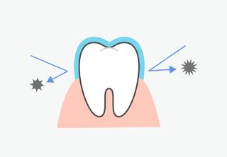
Periodontosis is a dystrophic disease of the periodontium, the structures that support the teeth. This complex pathological process disrupts cellular metabolism. The gums and the periodontal ligament do not receive enough oxygen and nutrients. This leads to the dystrophy of the periodontal tissues around the tooth. It changes the shape of the gums, reduces the height of the alveolar bone, and begins gum recession.
Dentists claim that the diagnosis of "periodontosis of the teeth" does not exist. In most cases, about 98%, patients are diagnosed with generalized aggressive periodontitis. The issue is that periodontosis develops unnoticed and painlessly. People seek medical attention too late, by which time hygiene problems and gum inflammation have already begun. The doctor diagnoses "periodontal disease" and starts comprehensive treatment.
Differences between Periodontitis and Periodontosis
Let's examine the differences between these terms:
- PeriodontOSIS — indicates a non-inflammatory pathological condition leading to dystrophy or changes in cell structure. In periodontosis, the nutrition of the tissues is disrupted, leading to gum reduction due to recession.
- PeriodontITIS — is an inflammatory disease. It is usually caused by bacteria, viruses, or other parasites. It leads to functional disturbances in the organs. Typical for periodontitis are the presence of plaque, subgingival tartar, formation of periodontal pockets, inflammation, and bleeding of the gums. The inflammation progresses, pockets enlarge, and teeth start to loosen.
| Periodontitis | Periodontosis | |
|---|---|---|
| Signs of disease | The periodontal pockets around the teeth are enlarged. Frequent bleeding and pus discharge from the gums. Teeth start to loosen in the early stages. | Occurs without inflammation and bleeding. Teeth become visible up to the neck and root. Teeth become mobile only in advanced stages. The gums recede. |
| What can I see on an x-ray? | Vertical bone resorption is visible, the bone becomes looser. Deep clinical pockets are present. | Horizontal bone resorption is visible. The bone tissue becomes denser. |
| Carious deposits | Soft plaque with dental calculus around the gums. | Dental calculus is often absent but can be large and hard above the gum line. |
| Rate of development | Rapidly progressing inflammation affects the gums and can lead to tooth loss. | The disease develops slowly and without symptoms. |
Symptoms of Periodontosis
How to identify periodontosis:
- Increased tooth sensitivity.
- No inflammation, bleeding, or swelling.
- Itching or burning in the gums.
- The mucous membrane is pale, interdental papillae are densified.
- Gum recession: Gums reduce in size, the neck or root of the tooth becomes visible.
- Wedge-shaped defect: Erosion of enamel near the tooth root.
At the first signs of illness, immediately schedule a consultation with a dental therapist. It's difficult to diagnose gum problems on your own, and without professional help, there's a risk of exacerbating the situation, including tooth loss.
Causes of Periodontosis
Periodontosis starts with the thickening of vessels and capillaries. This leads to poorer blood circulation, disrupting the supply of oxygen and microelements to the tissues. The tissues of the periodontium become denser, and the bone around the teeth decreases. Dystrophy causes gum recession and the loss of connection of soft tissue, periosteum, and ligaments to the tooth.
The structures of the gum transform into fibrous fibers (collagen tissue). The bone gradually atrophies, the height of the gum decreases, exposing the tooth root.
Other causes of periodontosis include:
- Harmful habits – smoking and excessive alcohol consumption, leading to dystrophy of the periodontal tissues.
- Incorrect bite – leads to increased chewing load and disrupts the blood supply to the tissues around the teeth.
- Nutritional imbalance – disrupted balance of proteins, fats, and carbohydrates or poor absorption of elements, incorrect diets without medical consultation.
- Hormonal changes (adjustments) – related to the endocrine system, changes in the thyroid gland, menopause in women, the natural aging process.
The jaw system has a certain resilience. This is similar to the automatic tire inflation system. If a tire is punctured, you have time to reach a service station to prevent further damage. The same applies to teeth: at the first signs of problems, local measures can be taken. If delayed, the problem will worsen, and comprehensive gum treatment or even surgical intervention may be required.
Classification and Stages
- The type of dental disease - dystrophy, causes changes or destruction of the structures around the teeth.
- The spread of the disease - generalized, affects the entire jaw (often both).
- Duration - more than 3 months, only in chronic form. There can be a remission phase (reduction of symptoms).
Stages of Periodontosis Development
1. Mild Stage:
Reduction of the alveolar ridge, a third of the tooth root is exposed.
Symptom-free, common complaints include itching, discomfort, or burning in different areas of the jaw.
Clinical signs: tension and functional insufficiency of the vessels in the periodontium. Pale gums, dense tissues around the teeth, visible tooth neck (recession up to 3 mm). X-ray shows initial bone atrophy, possible venous congestion.
2. Moderate Stage:
- Reduction of the interdental septa to half the root length.
- More noticeable symptoms: increased spacing between teeth, sensitivity to temperature changes, densification of the mucous membrane, numbness of the gums, accumulation of dental deposits, generalized gum recession of 3-5 mm, wedge-shaped defects.
3. Severe Stage:
- Atrophy of the alveolar ridge more than half the root length.
- Mobility of teeth, divergence in the frontal area.
- Gum recession of more than 5 mm, densified edge.
- Possible tooth loss.
In the late stages, periodontosis can be complicated by inflammation. X-rays reveal atrophy of the alveolar process and symptoms of periodontitis (bone resorption, deep periodontal pockets). Externally, the inflammation manifests as bleeding, abscess formation, and pus discharge. Surgical intervention may be required to preserve the teeth.
Treatment of Periodontosis
The treatment is usually complex, so one should not expect a quick cure after a single visit. There is no standard treatment plan – each case requires an individual strategy and treatment scheme. In some cases, improving personal oral hygiene is sufficient, while in complex cases, surgical periodontology, flap surgeries, open curettage, gum plastic surgery, splinting, etc., may be necessary.

Diagnosis
1. Anamnesis and complaint collection. The doctor conducts a thorough examination, inquires about previous dental diseases, chronic illnesses, and harmful habits. He identifies the causes and symptoms to determine indications and contraindications for treatment.
2. Oral examination. The doctor examines the jaw to determine the extent of the pathology, detect dental deposits and pathological tooth wear. He evaluates the color and density of the soft tissues and the anatomical bite. Using a probe, he measures the depth of the periodontal pockets.
3. Instrumental diagnostics:
- Biomicroscopy: Using a biomicroscope and fluorescent light, the doctor examines the capillary network of the periodontium and assesses the condition of the vessels and blood circulation.
- Rheoparodontography: Evaluates the condition of the vessels in the early stages of the disease.
- Polarography: Measures the oxygen balance in the tissues of the periodontium, checks circulation and oxygen absorption.
4. Radiological examination: Panoramic X-ray or CT to determine the stage of periodontosis, identifies osteosclerosis, clinical gum pockets, recession, and reduction of interdental septa.
Sometimes diagnosing a disease can be challenging. For example, X-rays may not show periodontological pockets, everything points to periodontosis, but the mucous membrane is inflamed. This can be due to poor oral hygiene. Accumulation of soft plaque and dental calculus can cause gingivitis (and potentially periodontitis). Comprehensive oral hygiene followed by treatment of periodontosis is necessary. However, if clinical pockets are present, more complex treatment is required.
Comprehensive Treatment in Dentistry
- The general treatment aims to correct microcirculatory disorders. Medications are used to improve blood circulation and clean the vessel walls, as well as to enhance metabolic processes.
- Local treatment includes oral sanitation: removal of plaque and calculus, elimination of inflammation. Professional oral hygiene is performed.
- Elimination of supercontacts involves selective grinding of the teeth. In periodontosis, calcium accumulates in the hard tooth structures, leading to a traumatic bite. The dentist corrects the bite and eliminates wedge-shaped defects.
- Orthodontic treatment is prescribed for severe bite anomalies.
- Symptomatic therapy includes remineralization of the teeth, temporary splinting for tooth mobility. Glass fiber or artificial crowns are used to correct tooth position and eliminate interdental spaces.
Always consult with your dentist about the timing and dosage of medications. Treatment should be carried out under medical supervision. Do not diagnose yourself after reading medical articles; it's better to schedule an appointment with a specialist.
Surgical Treatment Methods for Periodontosis
Before surgery, oral sanitation and removal of plaque are necessary. Depending on the condition of the gums, local anesthesia and surgical procedures are performed:
- Gingivoplasty (flap surgeries). Used to correct the height of the gum edge, cover exposed tooth roots, and hide wedge-shaped defects. Under anesthesia, a cut is made in the soft tissue, a flap is formed, and curettage is carried out.
- Curettage. This method is used for inflammation and the formation of periodontal pockets. Treatment of the tissue around the teeth with an ultrasonic scaler, antiseptic treatment, and removal of hard granulations. After the procedure, the gum is pressed against the tooth, and follow-up examinations are conducted.
Medication Treatment of Periodontosis
1. Reducing Tooth Sensitivity:
- Fluoridation of enamel in the dental office.
- Desensitizers to reduce sensitivity, seal the dentin canals.
2. Improving Metabolic Processes:
- Ascorutin for tissue regeneration.
- Vasodilators to improve microcirculation.
- B vitamins for carbohydrate, amino acid, and protein metabolism.
- Vitamin C combined with vitamin P to densify vessel walls.
3. Antibiotics:
- Used only in complicated periodontosis with inflammation.
4. Tooth Cleaning in Periodontosis:
- Mouthwashes with antioxidants to oxygenate sclerosed vessels.
- Fluoride-containing toothpastes with calcium and phosphorus, propolis-based pastes.
Discuss with your doctor the choice of dental products, including the specifics of taking fat-soluble and water-soluble vitamins. Do not take antibiotics without a doctor's prescription and in the wrong dosage! Find out how long you can use medicinal toothpastes.
Nutrition
Dentists recommend following a specific diet for periodontal problems:
- Avoid acidic foods (berries, fruits, juices) as they affect the sensitivity of tooth enamel. Reduce the consumption of sweets and carbonated drinks.
- Eat fresh fruits and vegetables. They clean teeth like a car wash, massage the gums, and provide the body with vitamins and macronutrients.
- Don't forget about seafood, nuts, and avocado. These foods contain unsaturated fatty acids that influence cellular metabolism.
- Add dairy products (cottage cheese, classic yogurt, cheese) to your diet. They are a good source of calcium and phosphorus, helping to restore hard tissues and enamel.
Consult a nutritionist for a balanced diet. They will help you find the right nutrition plan for your health condition.
Physiotherapeutic Procedures
- Electrophoresis: The treatment includes 10 sessions at weekly intervals. Heparin is used to normalize oxygen levels and improve blood circulation. A heparin-soaked bandage is applied to the alveolar ridge and fixed with electrodes. The procedure lasts up to 15 minutes.
- Vacuum Massage of the Gums: The vacuum device attachment is applied to the mucosa at several points. The pressure massage stimulates blood circulation and metabolic processes. A course of 10 treatments with five-day intervals is prescribed.
- Laser Therapy: Laser biostimulation accelerates healing and tissue regeneration. It reduces sensitivity in wedge-shaped defects and eliminates inflammation. Recommended for early and middle stages of periodontosis. Up to 15 sessions per year are possible.
Prognosis
The success of treatment depends on early diagnosis and treatment. The earlier the treatment starts, the higher the chances of a positive outcome. In advanced stages of periodontosis, bone atrophy and tooth loss may occur. In such cases, prosthetics or jaw implantation may be required.
Prevention of Periodontosis
Basics of individual prevention of periodontosis:
- Regular dental visits for comprehensive oral hygiene.
- Avoid bad habits, maintain a daily routine, and avoid stress.
- Regular and correct tooth brushing with medium-hard brushes and additional hygiene products.
- Daily finger gum massage, possibly using propolis gel. Do not massage during inflammation.
Preventative measures will not help cure periodontal disease at home. If pathology is present, timely treatment is required!
Who treats periodontosis?
A periodontologist is a specialist who diagnoses and treats problems of the periodontium (gums and bone around the teeth). He also performs procedures to strengthen the health of the maxillary structures and maintain the gum-tooth connection.
The periodontologist develops a treatment strategy and treats diseases of the surrounding gum tissue, such as:
- Periodontitis - infectious lesion of the periodontal tissue.
- Stomatitis - inflammation of the mucous membrane.
- Idiopathic anomalies - when the cause of the disease is unknown.
- Gingivitis - inflammation of the gums without destruction of the periodontal ligament.
- Periodontosis - non-inflammatory pathology, atrophy of the alveolar bone, recession of the mucous membrane.
The specialist also conducts preventive examinations, thorough cleaning (curettage) of the gum pockets from hard plaque, as well as professional teeth cleaning.
Frequently Asked Questions
What causes periodontal diseases?
The primary cause of periodontosis is the accumulation of dental plaque due to inadequate brushing and flossing. Other contributing factors include smoking, hormonal changes, certain diseases (such as diabetes), medications causing dry mouth, genetic predisposition, and poor nutrition.
How can periodontal diseases be prevented?
Prevention involves adhering to oral hygiene guidelines such as: brushing teeth twice a day, regularly using dental floss, using fluoride mouthwashes, undergoing dental exams and cleanings at least twice a year. Healthy eating and quitting smoking also significantly reduce the risk.
Can children suffer from periodontitis?
Although rare in children, they can still be at risk, especially if they develop poor oral hygiene habits. Teaching children the importance of regular tooth brushing and flossing is vital to prevent periodontitis in the future.
Is periodontal disease linked to other health problems?
Yes, periodontal diseases are associated with a number of other health issues, including heart disease, stroke, and diabetes. It's not just an oral health issue; it can significantly impact overall health, highlighting the importance of prevention and treatment.
