At Dental Educ, we discuss the importance of using microscopes in dentistry. These optical devices enable dentists to perform highly accurate diagnostics, significantly improving treatment quality. Microscopes assist in treating cavities, root canals, implantations, and tooth restorations. With their help, dentists can avoid medical errors and reduce the patient’s radiation exposure. The advantages of treatment under a microscope include high accuracy, gentle care, and quick recovery after procedures. Discover why this method is becoming increasingly popular among professionals and patients alike.
Treatment Under a Microscope – What Is It and Why?
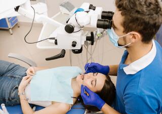
During dental treatment under a microscope, the dentist doesn’t lean over the patient; instead, they observe through the eyepiece. The electronic optical equipment is positioned over the surgical field using a pantograph, allowing the microscope to be easily and precisely aligned over the working area. The optical lens is about 25 cm above the patient’s head.
The dental microscope allows for taking photos and recording videos. The image from the built-in camera is transmitted to the assistant’s screen, enabling them to monitor the procedure and provide the dentist with the necessary instruments and materials.
This optical device ensures procedures like canal filling or tooth preparation are carried out under close visual control, significantly enhancing the quality of the results.
Using a microscope enables dentists to make precise diagnoses, reduce the need for X-rays, and eliminate the chance of unfortunate medical errors. Modern optics can even save a tooth that was deemed untreatable in other clinics.
Indications for Dental Microscope Treatment
- Diagnosing cavities and cracks in tooth enamel
- Removing decay, filling, and cosmetic restoration of teeth
- Endodontic treatment (for pulpitis, periodontitis, trauma, etc.)
- Strengthening a damaged tooth before prosthetics
- Implantation, prosthetics
- Surgical procedures for cysts, granulomas, etc.
Features of Treatment Using a Microscope
The scope of optical instruments in dentistry is vast and not always immediately obvious. They are used in therapy, including endodontic treatment, surgery, periodontology, orthopedics, and implantology.
During Root Canal Treatment
Optics allow for the detection of narrow canal openings. Working with 40x magnification of the localized area helps the dentist thoroughly clean the canals of affected tissue, stop inflammation, and properly fill the cavity. The microscope is invaluable for retreating root canals, enabling the careful removal of old filling material, dental posts, or broken instruments.
During Cavity Treatment
Using a microscope, the dentist can magnify the cavity 40 times, carefully removing the decayed tissue without damaging the healthy tissue. With this magnification, it becomes easier to avoid contact with the tooth pulp and prevent additional procedures. When placing a composite filling under a microscope, the dentist ensures the precise layering of the filling material. After polishing, the dentist inspects the joint between the filling and the tooth. The lens reveals even the smallest imperfections and roughness.
During Implantation
Tooth implantation typically involves tools visible to the naked eye. However, in complex cases—such as placing a titanium root near the maxillary sinus—the use of optics is justified. The device enhances the surgeon’s view of the surgical field, and the high magnification allows for an accurate assessment of the implant’s placement quality.
During Restorations
The use of 40x zoom enables the dentist to restore a tooth with maximum accuracy and recreate the lost anatomy of the tooth structure. When placing veneers, the optical device helps the dentist perform highly precise preparation and flawlessly secure the veneers in the smile zone.
Doctor's Advice
For some patients, the question arises whether a microscope is necessary during dental treatment. It’s important to note that there are no contraindications to the use of optical equipment, as it does not come into contact with tissues. As for its necessity, there are no diseases in therapy that cannot be treated without a microscope. However, the involvement of such equipment significantly improves the quality of the treatment outcome. A skilled dentist will always properly assess the need for optical assistance in your specific case.
Advantages of Dental Treatment Under a Microscope
- High accuracy in diagnosis. Using a microscope, the dentist can identify small cracks, wear and tear on old fillings, compromised sealing, and early-stage cavities.
- Gentle treatment of teeth. With a microscope, the dentist can see the boundary between healthy and damaged tissues, removing only the inflamed parts.
- Collaboration with other specialists. The dentist monitors the procedure on a computer screen, allowing them to consult with other experts if needed.
- Reduced recovery time after surgery. The focused action on damaged tissues only helps minimize recovery periods.
- Lower radiation exposure. Using a microscope reduces the need for frequent X-rays, as treatment progress can be monitored via the built-in camera at any stage.
- Lower risk of medical errors. Enhanced precision and control over procedures help minimize mistakes.
- Ability to save severely damaged teeth. When retreating root canals, the dentist can delicately reopen the canal, locate the issue, and correct it, preserving the tooth's functionality. Microscopes increase the chances of saving severely deteriorated teeth.
- Faster procedures. With optics, the dentist spends less time on canal fillings, cyst removals, and other treatments.
Frequently Asked Questions
For which procedures is a dental microscope used?
A microscope is used for diagnosing cavities, root canal treatments, restorations, implants, prosthetics, and surgeries such as cyst and granuloma removal.
How does a microscope improve cavity treatment?
A microscope magnifies the cavity 40 times, allowing the dentist to carefully remove decayed tissues without damaging the healthy ones, improving the quality of the filling.
What problems can a microscope solve during root canal treatment?
It helps locate narrow canal openings, thoroughly clean the canals, and precisely fill the cavity, preventing inflammation.
What are the indications for using a microscope in dentistry?
Indications include diagnosing cavities and enamel cracks, endodontic treatment, aesthetic restorations, implantations, prosthetics, and surgeries.
Are there any contraindications to using a microscope in dentistry?
There are no contraindications, as the microscope does not touch the patient's tissues. Its use depends on necessity and can greatly enhance treatment outcomes.
